Our patient today was healthy as far as he knew. However, he started feeling a lump on the side of his pelvis, right above his hip.
First, it was a very tiny subtle lump, so he didn’t mind.
With time, it started growing slowly. He wouldn’t notice any changes on a weekly basis, but after a year he could tell it was larger than in the beginning. Anyway, it didn’t hurt him or bother him so he didn’t do anything about it.
Finally, the lump was too large to ignore: you could see it even through his trousers. That’s when he decided to go the doctor.
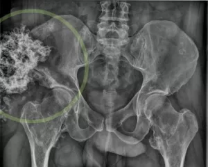
His doctor decided to perform an x-ray, to begin with. Here it is:

You can see there is something wrong, right? There is a large mass above his hip, and it “looks” like bone. It is a tumor with calcifications.
This raised the suspicion that it might be an aggressive tumor, so we performed several more tests, like an MRI and CT scan.
Even though this was the largest tumor, we found the patient had many small tumors in other bones. He had, we thought, a osteocondromathosis. It’s a disorder where you get many bone tumors. Fortunately, most of them are benign.
But what about the large tumor?
We performed MRI and CT scans that were like this:

It was large, there were many calcifications and it was attached to the pelvic bone.
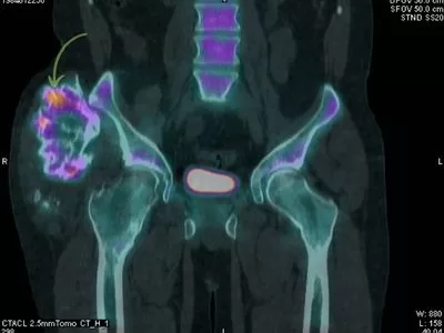
Also, the patient got a PET-CT, which tells us about its behavior: if it is “bright” it’s probably aggressive:

There was a small bright area on the upper side, so doctors decided it was best to remove it.
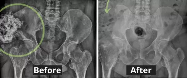
It was not an easy surgery but fortunately, it was not affecting any vital structures. They had to remove some of the iliac bone, but the hip joint was intact, so that didn’t cause him any trouble walking or standing.
You can see the before-after x rays:

After surgery, the patient recovered ok and didn’t have any main complaints. He is having MRI check-ups, just to make sure the tumor won’t come back.
Leave a Reply